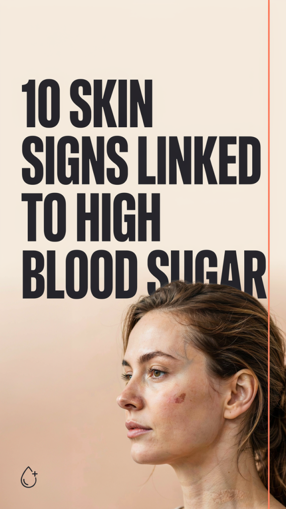
10 Skin Signs Linked to High Blood Sugar: What Your Skin May Be Telling You in 2026
Our skin often gives early clues about internal health, and high blood sugar is one of the conditions that shows up in surprisingly visible ways. In this guide we’ll walk through 10 skin signs linked to high blood sugar, explain the mechanisms behind them, and tell you what to watch for so you can act early. Whether you have diabetes, prediabetes, or just want to be vigilant, understanding these skin changes helps us catch problems sooner, reduce complications, and guide conversations with clinicians. We’ll balance practical takeaways (when to see a doctor, basic home care) with enough medical detail to make the signs memorable. Let’s start with how elevated glucose affects the skin at the cellular level, knowing the “why” makes the “what” easier to spot.
How High Blood Sugar Affects Skin: The Key Mechanisms To Know
High blood sugar alters skin health through several overlapping mechanisms. First, glycation, the non-enzymatic binding of glucose to proteins, stiffens collagen and elastin. That reduces skin elasticity and impairs barrier function, making skin drier and more fragile. Second, microvascular damage from chronic hyperglycemia narrows and stiffens small blood vessels, which reduces tissue perfusion and oxygen delivery: poor circulation slows repair and predisposes to ulcers. Third, immune dysfunction is common with elevated glucose: white blood cell chemotaxis and phagocytosis are less effective, so bacteria and fungi can multiply on skin that would otherwise resist infection. Fourth, neuropathy secondary to hyperglycemia leads to reduced sensation. Patients may not notice minor trauma that later becomes a persistent wound. Finally, lipid metabolism can be disturbed, producing deposits that show up as xanthomas or waxy plaques.
These mechanisms explain why a range of skin problems, from dark velvety patches to painless blisters, can be markers of high blood sugar. Recognizing the pattern (metabolic changes + vascular problems + impaired immunity) helps us interpret individual findings and decide whether they point to prediabetes, poorly controlled diabetes, or another condition that requires evaluation.
Acanthosis Nigricans — Dark, Velvety Patches Around Neck And Folds
Acanthosis nigricans is one of the most recognizable skin signs linked to high blood sugar and insulin resistance. Clinically it appears as symmetric, velvety, hyperpigmented patches most often on the posterior neck, armpits, groin, and under breasts. The texture is important: unlike simple tanning, these areas feel thickened and sometimes slightly raised. The underlying driver in many people is hyperinsulinemia: insulin (and insulin-like growth factors) stimulates keratinocyte and dermal fibroblast proliferation, producing the characteristic appearance.
We should note that acanthosis nigricans can vary with age and ethnicity, it’s more obvious in darker skin but also present in many lighter-skinned patients. It’s a red flag for metabolic syndrome: when we see it in clinic, we often recommend checking fasting glucose, HbA1c, and other cardiometabolic markers. Treatment focuses on addressing the root cause (weight loss, improved glycemic control, managing insulin resistance). Topical keratolytics, retinoids, or procedures like laser therapy can improve appearance but won’t replace metabolic treatment. If it appears suddenly and is extensive, we also consider, albeit rarely, paraneoplastic causes and investigate appropriately.
Diabetic Dermopathy — Shiny Brown Scars And Light Spots On The Shins
Diabetic dermopathy presents as round or oval, brownish, atrophic-appearing patches, usually on the shins. Patients often describe them as “shiny” or like old scars. These lesions are common in people with long-standing diabetes and are thought to result from minor trauma combined with microangiopathy: reduced dermal blood flow and structural collagen changes lead to localized skin breakdown and poor repair.
They’re typically asymptomatic and don’t require aggressive treatment, but they’re clinically useful. We use them as visual evidence of chronic microvascular disease and often reassess vascular risk factors when we find them. Management emphasizes optimizing blood sugar control and skin care: daily gentle cleansing, regular emollient use to prevent cracking, and sun protection to limit pigmentary change. If the lesions are symptomatic, expanding, or have atypical features (rapid growth, ulceration), we biopsy to rule out other causes such as stasis changes, necrobiosis, or even cutaneous malignancy.
Necrobiosis Lipoidica — Red‑Brown, Waxy Plaques That Can Thin Skin
Necrobiosis lipoidica is less common but more distinctive: it starts as well-demarcated red-brown papules that evolve into yellowish, waxy plaques with a glassy, atrophic center. The shins are the classic site. While the condition is associated with diabetes, not everyone with necrobiosis lipoidica has diabetes, yet the two share underlying vascular and collagen degeneration processes linked to abnormal glucose metabolism.
Patients often notice an initial bruise-like patch that slowly enlarges over months to years. The plaques can become thin and fragile, with telangiectasias and occasional ulceration after minor trauma. Treatment is challenging: topical and intralesional corticosteroids are first-line to reduce inflammation. For resistant cases we consider topical calcineurin inhibitors, systemic agents (like pentoxifylline or antiplatelet therapy), or phototherapy. Importantly, when we see necrobiosis lipoidica we review glycemic control and counsel on protective measures, shin guards, avoiding trauma, and prompt care for any breaks in the skin, because ulceration can be slow to heal in this setting.
Eruptive Xanthomas — Sudden Clusters Of Small Yellow Bumps
Eruptive xanthomas are sudden crops of small, yellow to flesh-colored papules, often with an erythematous halo. They most commonly appear on the buttocks, shoulders, and extensor surfaces. Unlike cholesterol-driven tendon xanthomas, eruptive xanthomas are linked to severe hypertriglyceridemia, which frequently coexists with poorly controlled diabetes.
The pathophysiology involves deposition of triglyceride-rich macrophages (foam cells) in the dermis when serum triglycerides spike. Clinically they’re alarming but usually benign and reversible: when we correct the lipid abnormality and improve glycemic control, the lesions resolve over weeks to months. Because eruptive xanthomas indicate significant metabolic derangement, we treat them as a systemic warning sign: check fasting lipids urgently, look for secondary causes (alcohol use, medications, hypothyroidism), and initiate lipid-lowering strategies and glycemic optimization. If present, they’re a cue for a broader cardiovascular risk assessment.
Recurrent Skin Infections — Why Bacteria And Fungi Thrive With High Glucose
People with high blood sugar are more prone to bacterial and fungal skin infections. Elevated glucose provides nutrient-rich surface secretions and weakens innate immunity, neutrophil chemotaxis, phagocytosis, and intracellular killing are all less effective in hyperglycemic environments. The result: more frequent boils, furuncles, cellulitis, and candidal intertrigo.
Fungal infections, especially candidiasis in skin folds and tinea in disrupted skin, are common. We often see recurrent or atypical presentations: persistent athlete’s foot, recalcitrant yeast infections under breasts or in groin, and protracted nail infections. Bacterial infections can be deeper and more severe: Staphylococcus aureus is a frequent culprit, and recurrent abscesses may reflect impaired host defense. Management includes targeted topical or systemic antimicrobials guided by severity, restoring skin barrier (gentle cleansers, emollients), and, crucially, improving glycemic control. For recurrent cases we also evaluate for additional immune-suppressing conditions and offer education on hygiene, prompt treatment of minor breaks in the skin, and footwear/nail care to avoid repeated trauma.
Slow Wound Healing And Diabetic Ulcers — Persistent Sores And Risk Factors
Slow wound healing is a hallmark complication of diabetes and one of the most serious skin-related concerns. Elevated glucose impairs every stage of the healing cascade: hemostasis, inflammation, proliferation, and remodeling. Add neuropathy (loss of protective sensation) and peripheral arterial disease (reduced blood flow), and you have the classic triad that predisposes to diabetic foot ulcers.
Ulcers typically occur on pressure points, plantar metatarsal heads, toes, and heel, but can appear anywhere. They start as callus, blister, or small sore and can progress to deep, infected wounds with exposed tendon or bone if not addressed. Our approach prioritizes prevention: routine foot inspection, daily moisturizing to prevent fissures (but not between toes), appropriate footwear, and smoking cessation. Once an ulcer develops, multidisciplinary care is essential, offloading pressure, debridement, infection control, vascular assessment, and optimizing glycemic control. Early referral to a wound clinic or podiatry service dramatically reduces the risk of progression to limb-threatening infection.
Dry, Itchy Skin (Xerosis) And Pruritus Linked To High Blood Sugar
Xerosis, abnormally dry skin, and generalized pruritus (itch) are common complaints among people with high blood sugar. Several factors converge: osmotic diuresis leads to dehydration, glycation alters barrier lipids, and small-fiber neuropathy can create itch sensations without obvious skin findings. Dry, cracked skin not only causes discomfort but also increases the risk of secondary infection.
Practical measures work well: we recommend regular liberal application of emollients with ceramides or petrolatum, lukewarm showers rather than hot baths, and mild, fragrance-free cleansers. For localized intense itch an antihistamine or short-term topical steroid can help, though chronically uncontrolled itching warrants evaluation for neuropathic itch or cholestatic/renal causes. Importantly, when dry skin coexists with other signs like recurrent infections or slow-healing wounds, it reinforces the need to check blood glucose and consider diabetes as an underlying contributor.
Diabetic Blisters (Bullosis Diabeticorum) — Painless Blisters That Appear Suddenly
Bullosis diabeticorum refers to spontaneous, noninflammatory blisters that occur in patients with diabetes. They usually appear on the hands, feet, and lower legs, are painless, and can be tense and large. The exact cause isn’t fully understood, but microangiopathy, glycation-related changes in the dermal-epidermal junction, and mechanical fragility from neuropathy all likely contribute.
Because the blisters are sterile and often heal on their own, management is conservative: protect the area from rupture and secondary infection, use sterile dressings, and monitor for signs of infection. If a blister is large or at high risk of breaking, careful aspiration under sterile technique can reduce tension while preserving the roof as a biologic dressing. We always evaluate for concomitant neuropathy and vascular disease when these blisters occur, and reinforce glycemic optimization. If infection is suspected, redness, purulent drainage, systemic signs, culture and appropriate antibiotics are indicated.
Conclusion
Our skin offers a window into metabolic health, and the 10 skin signs linked to high blood sugar we reviewed should prompt thoughtful evaluation rather than alarm. Some findings, acanthosis nigricans, eruptive xanthomas, necrobiosis lipoidica, point toward underlying insulin resistance or dyslipidemia, while infections, slow-healing ulcers, and diabetic blisters reflect impaired immunity and vascular damage from sustained hyperglycemia.
When we spot these signs, the next steps are clear: check glucose metrics (fasting glucose, HbA1c), assess cardiovascular and vascular risk factors, and engage multidisciplinary care when wounds or infections are present. Early recognition and better glycemic control not only improve skin outcomes but reduce long-term complications. If you notice any of these changes, document what you see, photograph progression, and bring it up with your healthcare provider, skin clues often give us the head start we need.