10 Signs Your Legs Could Reveal Poor Blood Flow — What To Watch For In 2026
Circulation problems in the legs are common, underrecognized, and potentially serious, yet many of us ignore early warning signs until they become emergencies. In this text we’ll walk through the top 10 signals your legs might be sending about poor blood flow, explain why they happen, and show what steps we can take to investigate and treat them. Our goal is to help you spot patterns early, understand whether symptoms point to arterial or venous disease, and know when to seek prompt medical care in 2026. This isn’t a replacement for professional advice, but a practical, evidence-informed guide so we all know what to watch for and why it matters.
How Blood Circulation Works In The Legs (A Quick Overview)
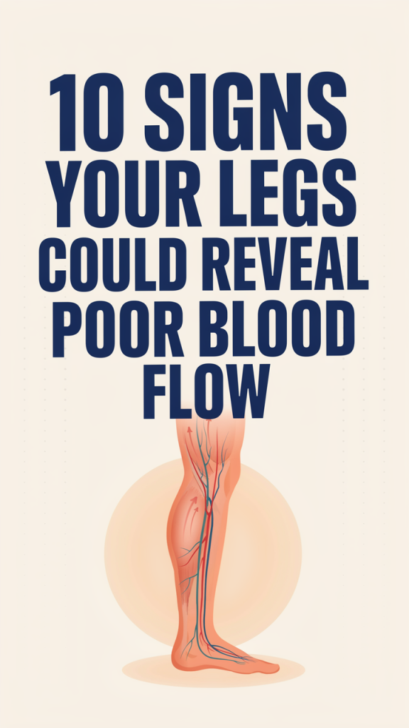
Blood circulation in the legs depends on two coordinated systems: arteries that deliver oxygen-rich blood from the heart to tissues, and veins that return oxygen-poor blood back to the heart. Arteries are high-pressure, muscular vessels: when they narrow from plaque (atherosclerosis) or spasm, downstream tissues get less oxygen and nutrients. Veins, by contrast, work with valves and muscle contractions to push blood upward. If valves fail or flow is sluggish, blood pools, causing different symptoms.
Why this matters: tissues with reduced arterial flow develop pain on exertion, coldness, and skin changes: tissues affected by venous insufficiency show swelling, discoloration, and ulcers from prolonged pressure and inflammation. Microcirculation (capillaries) also plays a role: diabetes, smoking, and chronic inflammation damage tiny vessels and slow healing.
Key risk factors we should keep in mind include smoking, diabetes, high blood pressure, high cholesterol, obesity, sedentary lifestyle, and age. Some conditions, like peripheral artery disease (PAD), primarily affect arteries, while chronic venous insufficiency (CVI) affects veins. Often they coexist, and distinguishing them matters because treatments differ: exercise programs and medications for PAD versus compression and venous procedures for CVI.
Pathophysiology snapshot:
- Arterial problems → reduced oxygen delivery → intermittent claudication (exercise pain), tissue pallor, cool skin, slow healing.
- Venous problems → increased venous pressure → edema, varicose veins, hemosiderin staining, venous ulcers.
Understanding these basics helps us interpret the signs described below and decide whether they point to arterial disease, venous disease, or another cause.
Persistent Leg Pain, Cramps, Numbness, Or Weakness With Activity
One of the most common and classic signs of poor arterial blood flow is intermittent claudication: aching, cramping, or burning pain in the calves (or thighs/hips) that starts with walking and eases with rest. We should pay attention when activity triggers consistent, predictable pain that forces us to stop and then improves after a few minutes. That pattern strongly suggests peripheral artery disease (PAD).
Nerve-related symptoms, numbness, tingling, or weakness, can overlap. With severe ischemia, muscles and nerves don’t get enough oxygen during activity, causing both cramping and sensory changes. It’s important to note differences: neuropathy (common in diabetes) can cause chronic numbness without activity-related triggers, while vascular claudication is tightly linked to exertion and relieved by rest.
Red flags and variations to watch for:
- Pain at rest (worse at night or when legs are elevated), more advanced arterial disease and demands urgent evaluation.
- Sudden severe pain with pale, cold limb, emergency sign of acute arterial occlusion.
- Pain that’s worse when walking uphill or fast, earlier-stage PAD typically affects calf muscles first.
What we can do: if we notice exertional leg pain that limits walking compared with peers, we should ask a clinician about an ankle-brachial index (ABI) test, a simple, noninvasive screening tool that compares blood pressure in the ankle and arm to detect arterial narrowing. Exercise programs supervised by a vascular specialist and risk factor control (stop smoking, optimize cholesterol and blood pressure, manage diabetes) are primary interventions. Medications like cilostazol can improve walking distance for some people, and revascularization (angioplasty or bypass) is reserved for more severe, lifestyle-limiting disease.
Coldness Or Noticeable Temperature Differences Between Legs Or Feet
When one leg or foot feels persistently colder than the other, poor arterial blood flow is a likely culprit. Because arteries supply warm, oxygenated blood, narrowing or blockage reduces warmth. We should be mindful of asymmetry: one cold foot compared with the other, or an overall persistent chill in both feet even though normal room temperature.
How to evaluate this at home: compare both feet by touch, feel the dorsum and sole, and note differences in temperature as well as skin texture. Check capillary refill by pressing a toenail or skin until it blanches and timing how long color returns, delayed refill (longer than 2 seconds) suggests compromised circulation.
Clinical context matters: cold feet can result from nonvascular causes (hypothyroidism, Raynaud’s phenomenon, or neuropathy), but if coldness accompanies other vascular signs, like pain on exertion, hair loss, or slow-healing wounds, arterial disease becomes more likely. Peripheral neuropathy can also cause a subjective sensation of cold without true temperature differences, so objective comparison and professional assessment are helpful.
When coldness is a serious sign: sudden onset of a cold, pale, and painful limb suggests acute ischemia and requires immediate emergency care. Chronic but progressive coldness with functional limitation warrants expedited outpatient vascular evaluation, ABI testing, and imaging if indicated.
Prevention and small steps: keeping active, quitting smoking, dressing warmly, and controlling cardiovascular risk factors help improve symptoms and slow progression. But persistent asymmetrical coldness should prompt clinical testing rather than home remedies alone.
Skin Color Changes, Shiny Skin, Hair Loss, And Nail Problems
Poor arterial perfusion causes characteristic changes in the skin and appendages because tissues aren’t getting enough oxygen and nutrients. We often notice:
- Pallor or a pale, waxy appearance when the leg is elevated.
- Dusky or cyanotic discoloration when dependent.
- Shiny, thin skin with visible venules and loss of normal creases.
- Hair loss on the legs and feet, and brittle or slow-growing toenails.
These signs develop gradually and may be subtle at first. Hair follicles and nail beds are sensitive to reduced blood flow: when supply is limited, growth slows and structures become fragile. Shiny skin and loss of elasticity are caused by chronic tissue undernourishment and atrophy.
Distinguishing features: venous disease often causes brownish discoloration (hemosiderin staining) around the ankles from chronic blood pooling, whereas arterial disease commonly produces a pale or bluish tone and pronounced skin thinning. Mixed disease is common in older adults.
Why this matters clinically: these cutaneous signs predict poor wound healing and increased infection risk. When we see hairless, shiny skin or nail dystrophy, we should consider a vascular assessment to measure perfusion and plan interventions. Early recognition allows us to prioritize revascularization or aggressive risk-factor modification before ulcers or gangrene develop.
Management focuses on addressing underlying vascular disease, meticulous skin care, and protecting feet from trauma. For people with diabetes, regular foot exams and podiatry involvement are especially important.
Slow-Healing Sores, Ulcers, Or Recurrent Infections On The Legs Or Feet
Nonhealing wounds on the legs or feet are a major red flag for poor blood flow. Wounds that linger for weeks, enlarge even though basic care, or recur after apparent healing suggest inadequate oxygen delivery and impaired immune function in local tissues.
Classic patterns:
- Arterial ulcers: typically on pressure points or areas with poor perfusion (toes, heel, lateral foot), often painful, with punched-out edges and a pale wound bed.
- Venous ulcers: usually around the medial ankle, accompanied by swelling and hemosiderin staining, often shallow with irregular borders.
Infection risk grows when circulation is impaired. Reduced blood flow limits delivery of immune cells and antibiotics: diabetic patients are at especially high risk because neuropathy may hide early injury and high blood sugar impairs healing.
Clinical approach: when sores are slow to heal we need a combined plan: evaluate arterial supply (ABI, toe pressures, duplex ultrasound), assess for infection (cultures, labs), and optimize wound care (debridement, moist dressings). For arterial-critical ischemia, revascularization, endovascular or surgical, often becomes necessary to restore perfusion and allow healing. Venous ulcers benefit from compression therapy and venous interventions when indicated.
Practical steps we can take: inspect feet daily, keep wounds clean, avoid smoking, control blood sugars, and seek prompt care for any sore that doesn’t show steady improvement within 2 weeks. Early vascular input makes a big difference for limb salvage.
Swelling, Varicose Veins, Or Sudden Leg Enlargement
Leg swelling is a common complaint and can reflect venous problems, lymphatic issues, or systemic disease (heart, kidney, or liver conditions). In the context of poor blood flow, chronic venous insufficiency (CVI) and varicose veins are frequent culprits. When venous valves fail, blood pools, venous pressure rises, and fluid leaks into tissues, causing edema and the visible bulging of varicose veins.
Symptoms and clues pointing to venous causes:
- Swelling that worsens by the end of the day and improves with leg elevation.
- Prominent, tortuous veins visible under the skin.
- A heavy, aching sensation in the legs, often relieved by compression or elevation.
But, swelling can also mask arterial disease, especially when both venous and arterial issues coexist. That’s why a careful assessment is essential: arterial disease may coexist with venous insufficiency, and compression therapy (standard for venous disease) can be unsafe if significant arterial compromise is present.
Also, sudden asymmetric enlargement of a leg, especially with pain and warmth, can signal deep vein thrombosis (DVT), a blood clot in the deep veins. DVT carries risk of pulmonary embolism and needs urgent evaluation.
We should hence evaluate swelling with a structured approach: history (timing, aggravating/relieving factors), physical exam (pitting vs nonpitting edema, skin changes, varicosities), and noninvasive testing (duplex ultrasound to assess venous reflux or clots: ABI to screen for arterial disease). Treatment ranges from conservative measures (compression stockings, exercise, weight loss) to procedures (endovenous ablation, sclerotherapy, valve repair) for symptomatic varicose veins, and anticoagulation for DVT.
Venous Versus Arterial Signs: How To Tell The Difference
Distinguishing venous from arterial problems is critical because management differs significantly. Here are practical contrasts we use in clinical assessment:
Onset and triggers:
- Arterial: pain with exertion (claudication), relief at rest, possible rest pain at night.
- Venous: aching or heaviness after standing, worse at day’s end, improves with elevation.
Skin and appearance:
- Arterial: pale, cool, shiny skin, hair loss, brittle nails, often distal ulcers on toes/pressure points.
- Venous: swollen limbs, brownish staining (hemosiderin) around ankles, varicose veins, shallow ulcers on the medial ankle.
Pulse and capillary refill:
- Arterial: weakened or absent pulses, delayed capillary refill.
- Venous: pulses usually present: issues are more with venous filling and reflux than arterial pulse.
Ulcer characteristics:
- Arterial: painful, deep, circular, minimal exudate.
- Venous: usually superficial, irregular borders, significant exudate and surrounding edema.
Diagnostic tests that clarify:
- Ankle-brachial index (ABI), first-line for suspected arterial disease.
- Duplex ultrasound, evaluates both arterial flow and venous reflux/clots.
- Toe-brachial index or transcutaneous oxygen pressure for patients with incompressible arteries (e.g., long-standing diabetes).
Why mixed disease matters: many patients have elements of both arterial and venous dysfunction. Compression is a mainstay for venous disease but must be used cautiously if ABI is low. Our role is to combine clinical clues with objective testing to create a safe, effective treatment plan tailored to the dominant problem.
When To See A Doctor — Tests, Urgency, And Common Treatment Options
Knowing when to seek medical attention can change outcomes. We recommend urgent evaluation for:
- Sudden severe leg pain, pallor, numbness, or inability to move a limb, possible acute arterial occlusion.
- Rapid swelling with pain and warmth, suspect deep vein thrombosis (DVT).
- Nonhealing wounds or ulcers that don’t improve within two weeks of basic care.
For less urgent but concerning symptoms (persistent exertional leg pain, chronic coldness, recurrent minor sores), schedule prompt outpatient evaluation.
Common diagnostic tests we rely on:
- Ankle-brachial index (ABI): easy, reliable screening for PAD.
- Duplex ultrasound: visualizes arterial stenosis and venous reflux or clots.
- CT angiography or MR angiography: used when planning revascularization.
- Angiography: gold standard for arterial mapping during interventional procedures.
- Blood tests: glucose, lipid panel, inflammatory markers as part of risk-factor assessment.
Typical treatment approaches:
- Lifestyle and risk-factor management: smoking cessation, supervised exercise therapy, blood pressure and lipid control, and diabetes management, these are foundational for both arterial and venous disease.
- Medications: antiplatelet therapy (aspirin, clopidogrel) for PAD, statins, anticoagulants for DVT, and medications like cilostazol for claudication in selected patients.
- Endovascular procedures: angioplasty with or without stenting to restore arterial flow: endovenous ablation for varicose veins.
- Surgical options: bypass surgery for severe PAD not amenable to endovascular repair: vein ligation or stripping in select venous cases.
- Wound care and adjuncts: compression for venous ulcers (after confirming adequate arterial supply), debridement, topical agents, and, when necessary, hyperbaric oxygen or advanced biologic dressings.
When we talk to patients, we emphasize a stepwise approach: confirm diagnosis with noninvasive testing, optimize medical therapy, and reserve interventions for symptomatic or limb-threatening disease. Multidisciplinary care, involving vascular medicine, interventional radiology, wound care, and primary care, often yields the best outcomes.
Conclusion
Leg symptoms are often windows into vascular health. By recognizing patterns, exertional pain, cold or pale feet, skin changes, slow-healing sores, swelling, and varicose veins, we can identify poor blood flow earlier and act before complications arise. Our best strategy combines vigilance, risk-factor control, timely testing (ABI and duplex ultrasound), and multidisciplinary treatment when needed. If you or someone you care for shows persistent or worrying leg symptoms, don’t wait: early evaluation improves mobility, quality of life, and can prevent limb loss. In 2026, with better screening and minimally invasive options, timely action makes a measurable difference.