10 Skin Signs That Could Reveal High Cortisol Levels (What To Look For in 2026)
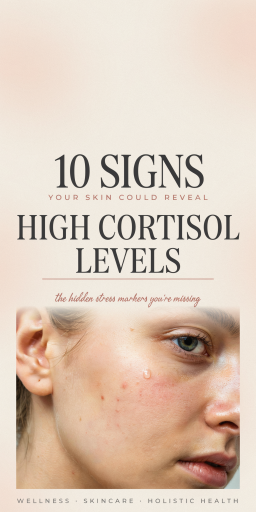
We often think of cortisol as “the stress hormone” and leave its effects confined to mood swings, sleep disruption, or weight gain. But cortisol shows up in our skin in very tangible ways, blemishes, thinning, slow healing, and subtle texture shifts can all be clues that our cortisol is chronically elevated. In this guide we’ll walk through ten skin signs that could point to high cortisol levels, explain why they happen, and offer practical next steps to investigate and address them. We won’t promise a miracle cure, instead, we’ll give clear, evidence-based reasoning and actionable tips you can use with your clinician or dermatologist in 2026.
How Cortisol Affects Skin: A Clear, Brief Overview
Cortisol is a glucocorticoid produced by the adrenal glands as part of the hypothalamic–pituitary–adrenal (HPA) axis response. Short-term surges are adaptive: long-term elevation remodels physiology. On skin, cortisol affects multiple cell types and signaling pathways: it alters keratinocyte proliferation, reduces collagen synthesis by dermal fibroblasts, changes immune surveillance, and shifts sebum production. The net result is a constellation of signs, thinner epidermis and dermis, reduced barrier function, impaired wound repair, and increased inflammatory responses that can present as acne, redness, or infections.
Two practical points we keep in mind: first, cortisol’s skin effects can come from endogenous overproduction (Cushing’s syndrome or chronic stress) or exogenous sources (topical/systemic corticosteroids). Second, cortisol interacts with other hormones, insulin, androgens, and estrogens, so skin findings rarely occur in isolation. That’s why a pattern of signs, rather than one single symptom, is more informative when thinking about cortisol.
In the sections that follow we’ll link each visible sign to the biological mechanisms so you can see why they make sense and what to do next. We’ll also highlight when a finding warrants urgent medical evaluation versus when it’s reasonable to start with lifestyle and skincare changes.
Acne Flares And Sudden Breakouts
One of the most common ways elevated cortisol shows up is through acne flares or sudden adult breakouts. Cortisol increases sebum production indirectly via stimulation of androgen precursors and sensitizing sebaceous glands. It also promotes low-grade inflammation by altering immune responses, neutrophil recruitment and cytokine expression go up, making pores more reactive.
Clinically, cortisol-related acne tends to appear as inflammatory, tender papules and pustules concentrated along the jawline, chin, and lower face in adults, though it can affect the forehead and back. Breakouts may coincide with periods of intense stress, sleep deprivation, or significant life changes. We often see patients report that their “stress acne” is not very responsive to over-the-counter benzoyl peroxide alone.
What to do:
- Track timing. Keep a simple log correlating stressors, sleep, and flare days, patterns are revealing.
- Combine topical approaches. Use evidence-based anti-inflammatories (topical retinoids, niacinamide, azelaic acid) plus targeted benzoyl peroxide for bacteria.
- Consider hormonal evaluation. If adult-onset or jawline-predominant acne accompanies other cortisol signs, testing cortisol levels or consulting endocrinology may be appropriate.
We should note that acne has many causes, diet, medications, and PCOS can mimic stress-related flares, so use the wider clinical picture rather than assuming cortisol is always to blame.
Thinning Skin, Stretch Marks, And Easy Bruising
Cortisol reduces collagen and elastin synthesis by inhibiting fibroblast activity and promoting matrix metalloproteinases that break down extracellular matrix. Over time, this leads to noticeable thinning of the dermis, skin that looks more translucent, fine-lined, and delicate. Along with reduced structural integrity, capillaries become more fragile, making bruises appear with minor trauma.
Stretch marks (striae) are a classic sign of prolonged cortisol excess. They commonly develop on the abdomen, flanks, thighs, and breasts and begin as pink or purple linear streaks that later fade to white-silver. When we see new, wide, and numerous stretch marks in a non-pregnant person, especially if accompanied by other red flags, we think about evaluating cortisol sources.
Practical tips:
- Protect and reinforce. Use topical retinoids (when safe), vitamin C serums, and peptides that support collagen synthesis: sunscreen is essential to reduce further photodamage.
- Avoid unnecessary topical steroids. Prolonged or high-potency topical corticosteroid use can mimic endogenous cortisol excess and accelerate thinning and bruising.
- Seek assessment. Sudden, widespread thinning, extensive striae, and easy bruising should prompt endocrine evaluation for endogenous hypercortisolism (Cushing’s) or review of prescribed corticosteroid use.
Remember: age and genetics also affect skin thickness, but the pattern and rapidity of change differentiate cortisol-driven changes from normal aging.
Corticosteroid Use Versus Endogenous Cortisol Effects
It’s important to distinguish exogenous corticosteroid effects from endogenous cortisol overproduction. Topical steroids, inhaled corticosteroids, and systemic prednisone can all produce skin findings similar to Cushing’s, including thinning, striae, delayed healing, and acneiform eruptions. Key clues we use:
- Distribution and timing: Topical steroid damage is often localized to treated areas but can generalize with potent or prolonged use. Exogenous systemic steroids produce a timeline tied to medication start and dose.
- Additional signs: Systemic steroids may cause weight gain, facial rounding (“moon face”), and proximal muscle weakness, similar to endogenous disease.
- Reversibility: Some steroid-induced changes improve after tapering or discontinuing exogenous steroids, though recovery varies.
If you’re using any corticosteroid product and notice these skin changes, we recommend discussing dose reduction or alternatives with the prescribing clinician before stopping abruptly. For suspected endogenous cortisol excess, coordinated testing (late-night salivary cortisol, 1-mg overnight dexamethasone suppression test, or 24-hour urinary free cortisol) should be pursued with an endocrinologist.
Slow Wound Healing And Recurrent Skin Infections
Cortisol suppresses many aspects of the immune response: it reduces lymphocyte proliferation, impairs macrophage function, and shifts cytokine profiles. On skin, this immunosuppression translates to slower re-epithelialization after cuts, delayed scab formation, and higher susceptibility to bacterial, fungal, or viral infections.
In practice we see wounds that take longer than expected to close, recurrent cellulitis in the same area, or persistent fungal tinea that won’t clear with standard topical therapy. Post-procedure healing (after minor dermatologic procedures or cosmetic treatments) may be prolonged or complicated by infections.
What to monitor and do:
- Track healing times. If a small cut or biopsy site remains open beyond the expected window (typically a couple of weeks for superficial wounds), get it checked.
- Optimize metabolic factors. High glucose impairs healing, so screen for diabetes when healing is poor.
- Reinforce barrier care. Gentle cleansing, topical antimicrobial dressings when indicated, and avoiding unnecessary topical steroids on infected skin help.
- Get specialist input. Recurrent infections or markedly delayed healing warrant an endocrine workup and infectious disease or dermatology referral.
We emphasize that delayed healing can also come from poor circulation, smoking, or nutritional deficits, so an integrative assessment is essential.
Increased Oiliness, Enlarged Pores, And Texture Changes
Many patients notice a change in skin texture when under chronic stress: pores look larger, the skin feels oilier, and overall texture becomes less refined. Cortisol elevates free fatty acid release and can indirectly upregulate sebum production. Over time, excess sebum plus impaired keratinocyte turnover clogs follicular openings and accentuates pore size.
Texture changes also stem from reduced collagen and altered hydration. We often describe this as a ‘‘grainy” or uneven feel, with light reflecting less smoothly. These changes are subtle at first but become more apparent with high-resolution selfies or when comparing recent photos.
Practical management:
- Start gentle exfoliation. Weekly chemical exfoliation (AHA or low-strength BHA) can improve texture and pore appearance: avoid over-exfoliating, which worsens barrier dysfunction.
- Use lightweight, non-comedogenic moisturizers with humectants (hyaluronic acid) to balance hydration without adding oil.
- Consider procedural options. For persistent texture and enlarged pores, microneedling or fractional lasers, performed when healing is adequate, can stimulate collagen and refine texture. Discuss cortisol or steroid status with your provider before procedures.
We remind readers that visible pore size has a genetic component: interventions improve the appearance but won’t eliminate pores entirely.
Facial Redness, Flushing, And Visible Capillaries
Chronic cortisol imbalance can contribute to vasodilation and fragility of small blood vessels, leading to persistent facial redness, flushing episodes, and telangiectasias (visible capillaries). Cortisol’s effects on connective tissue weaken perivascular support, making vessels more prominent and reactive to heat, alcohol, or stress.
When redness is new, diffuse, and accompanied by other cortisol signs, we consider a hormonal contribution. But, redness is multifactorial: rosacea, alcohol, spicy foods, sun damage, and certain medications also provoke vasodilation.
Management strategies:
- Identify triggers. Keep a diary of foods, beverages, medications, and stressors that provoke flushing.
- Calming topicals. Niacinamide, azelaic acid, and gentle barrier-supporting moisturizers reduce baseline inflammation: avoid sensitizing ingredients like high-concentration vitamin C or alcohol-based toners if skin is reactive.
- Procedural options. Intense pulsed light (IPL) or vascular lasers can reduce telangiectasias, but healing and response may be poorer if immune function or collagen support is compromised by cortisol issues.
If flushing coexists with systemic signs (weight change, muscle weakness, mood shifts), we’d escalate to endocrine testing. For isolated facial redness, dermatologic evaluation for rosacea or photodamage is usually the first step.
Dark Circles, Puffiness, And Under‑Eye Changes
Under-eye changes are a common cosmetic complaint and can be a window into systemic physiology. Elevated cortisol influences fluid distribution and promotes salt and water retention via mineralocorticoid receptor activation pathways, contributing to periorbital puffiness. Cortisol-driven thinning of periorbital skin reveals underlying vasculature and fat pockets, making dark circles more conspicuous.
We also see sleep disruption, often cortisol-related, exacerbate under-eye shadows: fragmented sleep increases inflammatory mediators and alters skin repair, magnifying pigmentation and pallor. In short, puffiness and dark circles are rarely caused by a single factor.
Practical steps:
- Improve sleep hygiene. Prioritize consistent sleep windows, reduce evening screen time, and limit late caffeine to address a common upstream driver.
- Topical brighteners. Low‑strength retinoids and vitamin C can improve pigmentation over months: caffeine-containing eye creams reduce transient puffiness by vasoconstriction.
- Medical evaluation. If puffiness is severe, asymmetric, or rapidly progressive, we’d consider thyroid function and renal/cardiac causes plus to cortisol assessment.
We advise patience: under-eye improvements from hormonal or sleep interventions can take several weeks to months to show meaningful change.
Conclusion
Our skin is a mirror of internal physiology, and cortisol leaves several recognizable fingerprints: acne flares, thinning, stretch marks, slow healing, oiliness, redness, and under-eye changes. When multiple signs cluster together, we should consider cortisol as a contributing factor and pursue a coordinated approach: track symptoms, optimize lifestyle (sleep, stress management, nutrition), review medications (especially corticosteroids), and consult dermatology and endocrinology when findings are sudden, severe, or progressive.
We recommend starting with small, measurable steps: keep a symptom-and-stress log for a few weeks, protect skin with sunscreens and barrier-supporting topicals, and prioritize restorative sleep. If you suspect true hormonal imbalance, simple screening tests and a specialist discussion can clarify next steps. By looking at the pattern rather than a single symptom, we can better decide when to self-manage and when to escalate care, and that practical distinction is often the fastest path back to healthier skin in 2026.