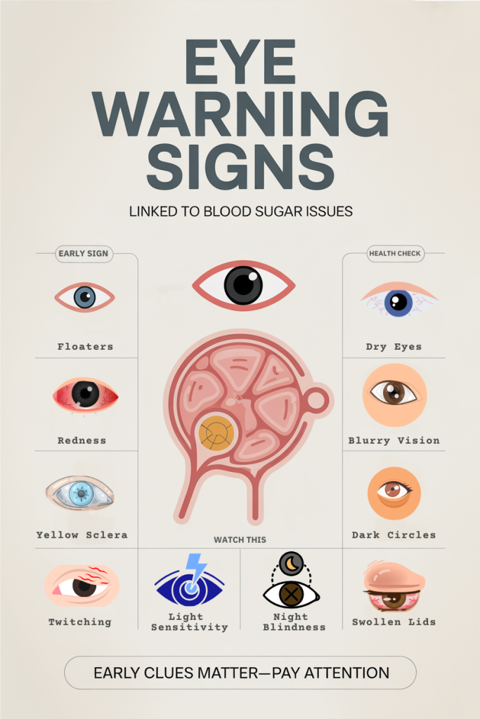
10 Eye Changes That Could Signal Blood Sugar Problems — What To Watch For
In 2026, as diabetes and prediabetes remain common but often underrecognized, our eyes can give early and actionable clues about blood sugar control. We’ll walk through ten specific eye changes that are frequently linked to high or fluctuating blood glucose, what they look like, why they happen, and when to act. Seeing a subtle blurring one morning or noticing new floaters doesn’t always mean diabetes, but when these symptoms appear together or persist, they can point to metabolic trouble that deserves investigation.
This article synthesizes current clinical understanding and practical advice so we can spot warning signs earlier. We’ll use plain language, real-world examples, and clear guidance on urgency and next steps, so you and the people you care for can get timely testing, treatment, or referral. Our focus is on what to watch for in daily life, how blood sugar affects eye structures, and when to seek emergency care or specialist input.
Blurred Or Distorted Central Vision (Macular Edema And Early Cataract Changes)
Blurry or distorted central vision often affects tasks that require fine detail, reading, recognizing faces, or seeing the center of a phone screen. When blood sugar is poorly controlled, two common mechanisms can produce these symptoms: diabetic macular edema and acceleration of cataract formation.
Diabetic macular edema occurs when fluid leaks from damaged retinal blood vessels into the macula, the central area responsible for sharp vision. Patients typically report straight lines appearing wavy, letters missing in the middle of words, or a gradual loss of central clarity. We should note that macular edema can be episodic, worse after periods of high blood glucose, and it’s a leading cause of vision loss in people with diabetes.
High blood sugar also alters lens proteins and hydration, which can hasten cataract development. Early cataract changes often cause generalized blurring, glare around lights, and difficulty with contrast, symptoms that overlap with macular disease, making professional assessment necessary.
What to watch for: new or worsening central blur, metamorphopsia (straight lines looking warped), increased glare at night, or a sudden need for stronger spectacles. We recommend prompt eye evaluation when these symptoms appear, ideally a dilated retinal exam and optical coherence tomography (OCT) if macular involvement is suspected. Timely treatment (anti-VEGF injections, focal laser, or cataract surgery when indicated) can preserve vision and is most effective when started early.
Vision That Fluctuates With Your Blood Sugar Levels
Fluctuating vision, where clarity improves and worsens across hours or days, is a hallmark sign that our eyesight is tracking blood sugar swings. When glucose levels rise, the lens inside the eye can absorb more sugar and swell slightly, temporarily changing its shape and refractive power. Conversely, when glucose falls, the lens returns toward baseline shape and our prescription may shift again.
People often notice they need different reading glasses from one week to the next or that distance vision is unexpectedly worse after a period of uncontrolled eating or missed medications. These refractive shifts usually reverse with stable glucose control over several weeks, but if fluctuations are frequent, they can interfere with daily activities and make accurate prescription correction difficult.
Practical tips: we should check blood sugar when vision changes acutely and avoid getting new glasses until glucose has been stable for several weeks. For clinicians, measuring point-of-care glucose and asking about recent glycemic variability should be part of the eye history. For patients on insulin or medications, patterns of large glucose swings warrant a discussion with primary care or endocrinology, stabilizing glycemia reduces refractive instability and the risk of more serious microvascular complications.
Floaters, Spots, Or Cobwebs — Signs Of Diabetic Retinopathy Or Vitreous Bleeding
New floaters, spots, or cobweb-like shadows passing through the visual field are often caused by particles within the vitreous gel or bleeding from retinal blood vessels. In people with diabetes, progressive retinal damage, diabetic retinopathy, can lead to fragile new blood vessels that bleed into the vitreous. This vitreous hemorrhage produces sudden floaters or a gray veil that may partly obscure vision.
The risk of vitreous bleeding rises with duration of diabetes, poor glycemic control, hypertension, and advanced retinopathy. Patients may describe sudden onset of many small spots or a dense cloud that reduces vision. In less severe cases, floaters come from benign posterior vitreous detachment, but in people with diabetes the distinction needs urgent evaluation.
When we see new floaters or a shower of spots, we treat it seriously: an ophthalmic exam with dilated fundoscopy is necessary. Imaging (B-scan ultrasound) helps when the view of the retina is blocked by blood. Management ranges from observation for small, clearing hemorrhages to vitrectomy surgery for dense, non-clearing bleeds or associated retinal tears. Importantly, optimizing blood sugar, blood pressure, and lipid levels reduces the chance of recurrent hemorrhage and slows retinopathy progression.
Dark, Shadowy, Or Missing Areas In Your Visual Field
Noticing dark patches, shadows, or missing sections in our peripheral or central vision can indicate localized retinal damage, often from diabetic retinopathy or ischemic events. These scotomas (blind spots) may start small and expand, or they can appear suddenly if a retinal vessel becomes blocked.
Diabetic retinopathy can create areas of nonperfusion where the retina doesn’t receive adequate blood flow. Those ischemic regions can cause function loss and prompt the release of VEGF (vascular endothelial growth factor), which in turn promotes abnormal vessel growth and leakage. The result: incremental blind spots, distorted vision, or, if central areas are involved, severe functional loss.
Another cause of sudden visual field defects is a branch retinal artery or vein occlusion, which is more common in people with vascular risk factors including diabetes, hypertension, and high cholesterol. Patients may report a curtain or shadow moving across their vision.
If we or our loved ones experience new areas of missing vision, we should seek ophthalmic assessment promptly. Visual field testing, dilated exam, OCT, and fluorescein angiography may be used to localize the problem and guide treatment. Early detection allows interventions, anti-VEGF therapy, laser, or systemic risk-factor optimization, that can prevent further vision loss.
Sudden Or Painless Vision Loss — When To Seek Emergency Care
Sudden or painless vision loss is alarming and can be caused by several eye emergencies that are more likely or more severe in people with diabetes. Examples include central retinal artery occlusion (CRAO), dense vitreous hemorrhage, advanced retinal detachment, or severe ischemic optic neuropathy. Because these conditions may lead to permanent damage if not addressed quickly, they’re true emergencies.
We should treat the following as reasons for immediate evaluation: a sudden curtain or shadow coming over part of the vision, abrupt loss of vision in one eye, or a rapid deterioration over hours. Some conditions, like CRAO, carry a very narrow window for intervention to preserve vision, and systemic evaluation for stroke risk is warranted.
Notably, painful vision loss can point to other issues such as acute angle-closure glaucoma or severe ocular infection, but painless loss is more typical of vascular or vitreoretinal causes. If you experience sudden loss, call emergency services or present to an eye emergency clinic, don’t wait for a routine appointment. While we act, ensuring blood glucose, blood pressure, and anticoagulation status are communicated to emergency clinicians helps guide acute management and reduces systemic risk.
Double Vision And Drooping Eyes From Nerve Damage
Diplopia (double vision) and ptosis (drooping eyelid) can occur when cranial nerves that control eye muscles are affected. Diabetes predisposes people to microvascular cranial nerve palsies, most commonly the third, fourth, or sixth nerve, leading to sudden, often isolated double vision. These palsies typically cause one eye to be misaligned, producing horizontal or vertical diplopia that’s worse when looking in specific directions.
Diabetic nerve palsies are often painless and may improve over weeks to months as ischemia resolves. But, a third nerve palsy that includes pupil involvement (dilated, unreactive pupil) raises concern for aneurysm and requires urgent neuroimaging. We should also remember that thyroid eye disease, myasthenia gravis, and brainstem lesions can cause similar symptoms: diabetes doesn’t exclude other diagnoses.
Evaluation includes careful ocular motility testing, pupillary exam, glycemic assessment, and often neuroimaging when atypical features exist. Management is mostly supportive, occlusion of one eye, prism glasses, or temporary patching, and addressing the underlying vascular risk factors. If diplopia persists beyond three months, strabismus surgery or botulinum toxin may be considered.
Poor Night Vision And Reduced Contrast Sensitivity
Difficulty seeing in low light or noticing that objects blend into backgrounds are signs of reduced rod function and impaired contrast sensitivity, changes that are common with diabetic retinal dysfunction. Even before clear retinopathy is visible on exam, subtle neural and microvascular changes can degrade how well the retina detects low-contrast or low-light stimuli.
Patients often report driving at night becoming harder, increased glare from oncoming headlights, or trouble navigating dim rooms. These deficits may be among the earliest functional complaints in people with metabolic risk and can precede clinically apparent retinopathy. They’re also important because they affect safety, night driving, stair navigation, and workplace tasks can become hazardous.
Testing for contrast sensitivity and scotopic (low-light) function isn’t routine in every clinic but can give useful baseline information. For us, recognizing complaints of night blindness should prompt a full retinal assessment, blood sugar evaluation, and counseling on safety adjustments. Maintaining stable glycemia, controlling blood pressure, and timely referral to ophthalmology can slow progression of these visual function deficits.
Red, Irritated, Or Chronically Dry Eyes Linked To High Blood Sugar
Persistent red, irritated, or dry eyes are frequently reported by people with diabetes. High blood sugar affects tear production and tear film composition, increasing the risk of meibomian gland dysfunction, ocular surface inflammation, and reduced corneal sensitivity. The result is often chronic dryness, burning sensation, and redness that don’t respond well to occasional artificial tears.
Diabetes also increases susceptibility to infections, such as bacterial keratitis, because of impaired corneal nerve function and delayed epithelial healing. Even minor corneal abrasions can heal slowly, prolonging discomfort and infection risk. In contact lens wearers with diabetes, vigilance is especially important.
Management combines symptomatic relief and addressing underlying causes: regular lubricating drops, eyelid hygiene, warm compresses for meibomian gland dysfunction, and topical anti-inflammatories when prescribed. Crucially, improving glycemic control often reduces severity. If redness is persistent, associated with vision changes, or accompanied by discharge, an eye care professional should evaluate for infection, corneal ulceration, or neurotrophic keratitis.
Eye Pain, Pressure, Or Headaches That May Indicate Glaucoma Or Optic Nerve Stress
While sharp eye pain and severe headaches often point to acute angle-closure glaucoma or inflammatory processes, subtler eye pressure sensations and recurrent headaches may reflect chronic optic nerve stress related to vascular disease. People with diabetes have an increased prevalence of glaucoma and may experience optic nerve ischemia from microvascular disease.
Open-angle glaucoma is often painless but can cause progressive peripheral vision loss over years: patients may notice difficulty seeing at the sides or bumping into objects. Acute angle-closure glaucoma, by contrast, is an emergency with severe eye pain, nausea, halos around lights, and markedly elevated intraocular pressure. Diabetes doesn’t directly cause angle-closure but increases coexisting vascular risk, making comprehensive assessment essential.
When we or our patients report persistent eye pressure, new headaches associated with visual changes, or colored halos around lights, measurement of intraocular pressure, optic nerve assessment, and visual field testing are warranted. Early detection of glaucoma preserves vision: controlling blood pressure, avoiding nocturnal hypotension, and ensuring regular ophthalmic follow-up are key strategies for prevention and management.
Conclusion
Our eyes can reveal metabolic trouble long before we feel systemic symptoms. The ten changes we described, blurred central vision, fluctuating sight, floaters, dark spots, sudden loss, double vision, poor night vision, chronic dryness, and eye pain or pressure, each have specific mechanisms tied to blood sugar or vascular health. Recognizing these signs prompts timely testing for diabetes or for complications in people already diagnosed.
We should act on new or persistent visual symptoms rather than dismissing them as “just getting older.” Simple steps, checking blood sugar, scheduling a dilated eye exam, and optimizing blood pressure and lipids, can make a big difference. If symptoms are sudden, painless loss of vision, a curtain-like shadow, or severe eye pain with nausea, seek emergency care immediately. By staying alert to these changes and coordinating care between primary care, endocrinology, and ophthalmology, we protect sight and overall health in 2026 and beyond.