10 Ways Your Eyes Reveal You’re Sleep Deprived (And What To Do About It)
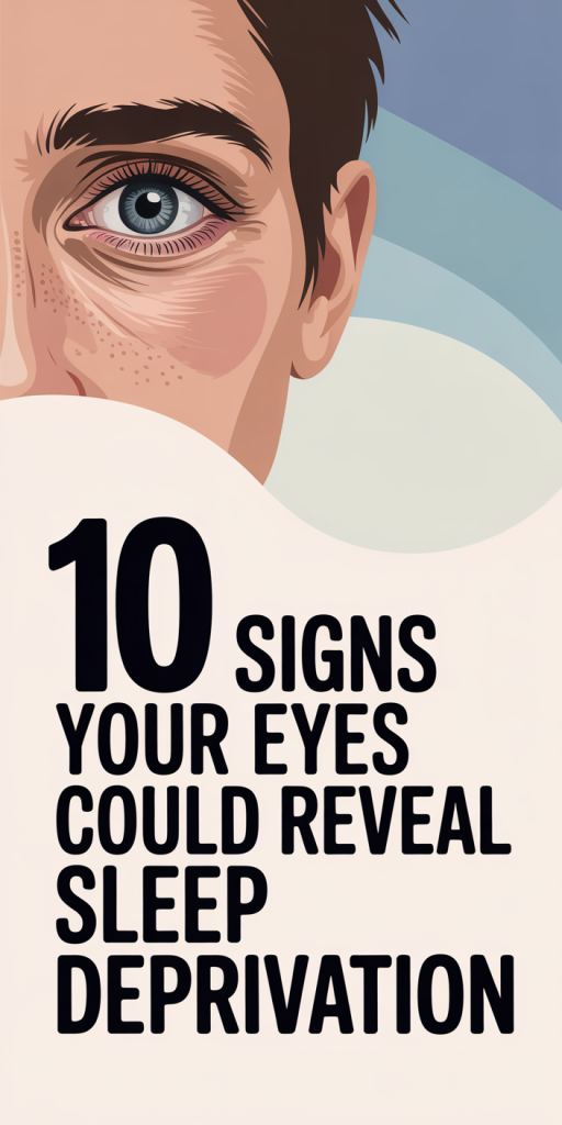
We’ve all had mornings when our reflection looks like we stayed up worrying all night, and sometimes, our eyes are the first clue that we didn’t get enough sleep. Eyes are remarkably sensitive to the effects of sleep loss: from tiny bloodshot veins to subtle changes in blinking and tear production, these signals give us early warnings about our overall rest and health. In this text we’ll walk through ten specific signs our eyes use to tell us we’re sleep deprived, explain the underlying causes, and give practical steps to ease symptoms or know when to get medical help. If you want to read your body like a book, start with the eyes.
How Sleep Loss Affects Eye Health And Appearance
Sleep plays a central role in restoring the body, and the eyes are no exception. When we don’t get enough restorative sleep, several physiological processes that protect ocular surface health slow down or become imbalanced. Tear film production and composition shift, eyelid muscles fatigue, and circulatory changes increase capillary permeability around the eyes. The result: more dryness, redness, puffiness, and slower reflexes.
Beyond the surface, chronic sleep deficit raises inflammatory markers and stress hormones that can worsen existing eye conditions like dry eye disease and blepharitis. Our skin around the eyes is thin and richly vascularized, so even small changes in blood flow or fluid balance become visible as dark circles or puffiness. Cognitively, sleep deprivation reduces our blink rate and the quality of blinks (they’re more incomplete), which leads to faster tear evaporation and fluctuating vision.
Put simply: eyes aren’t just cosmetic indicators, they reflect underlying physiological strain. Paying attention to ocular clues helps us catch sleep problems early and prevent complications like persistent dry eye, worsening refractive stability, or daytime visual impairment.
Red Or Bloodshot Eyes: Why They Happen And How To Spot Them
Red or bloodshot eyes are one of the most obvious signs that we’ve skimped on sleep. The whites of the eyes (sclera) normally appear slightly off-white because they contain small blood vessels. Sleep loss, especially when combined with rubbing, screen time, or environmental irritants, causes these vessels to dilate and become more visible.
Why it happens: sleep restriction increases sympathetic activity and inflammatory mediators that lead to conjunctival vessel dilation. At the same time, reduced tear production makes the ocular surface more vulnerable to irritants, which further provoke redness.
How to spot it: look for a diffuse pink or red appearance across the sclera rather than a localized area of redness. If the redness is accompanied by severe pain, a sudden decrease in vision, light sensitivity, or colored discharge, that suggests a more serious condition (like keratitis or acute glaucoma) and we should seek urgent care.
Quick relief tips: rinse with preservative-free artificial tears to rehydrate the surface and avoid rubbing the eyes. A cool compress for 5–10 minutes can constrict superficial vessels and reduce visible redness. We should also remove contact lenses and limit screen exposure until the redness subsides.
Dark Circles, Under‑Eye Hollows, And What They Mean
Dark circles are a common complaint and a classic sign of poor sleep, but they rarely have a single cause. In people who are sleep deprived, the skin under the eyes can appear darker for three main reasons: increased blood pooling in thin under-eye skin, changes in pigmentation, and shadowing from under-eye hollows.
Blood pooling and vascular visibility occur because sleep loss affects microcirculation and increases venous congestion around the orbital area. The skin under the eye is the thinnest on the body: when blood vessels dilate or blood pools, the bluish or purplish tint shows through easily.
Under-eye hollows deepen with fatigue because tissue-swelling patterns change: in some cases fluid accumulates in the eyelid during the night and redistributes when we’re upright, creating shadows. Chronic sleep deprivation can accelerate facial volume loss and make hollows look worse over time.
What to do: we can improve appearance by prioritizing regular sleep, elevating the head at night to limit fluid shift, and using cold compresses in the morning. Topical caffeine-containing eye creams or concealer help temporarily by constricting blood vessels and masking color, but restorative sleep is the most reliable fix. If dark circles are persistent even though good sleep and skincare, screening for allergies, anemia, or pigment disorders is sensible.
Puffy Or Swollen Eyelids: Fluid, Inflammation, And Timing Clues
Puffy eyelids are often blamed on late-night snacks, salt, or allergies, and while those factors matter, sleep deprivation itself shifts the body’s fluid balance and immune signaling in ways that create swelling.
Mechanisms: when we don’t sleep enough, cortisol patterns get disrupted. Elevated evening cortisol and altered aldosterone can encourage fluid retention. At the same time, inflammatory cytokines rise, increasing vascular permeability and allowing more fluid into the eyelid tissues. Incomplete blinking and reduced lymphatic clearance during poor sleep further impede the normal overnight removal of interstitial fluid.
Timing clues help us differentiate causes: transient morning puffiness that improves after a cold compress or gentle massage often points to fluid redistribution from poor sleep. Puffiness that’s consistent all day, itchy, or associated with redness may indicate allergies or blepharitis. Sudden, painful swelling with redness warrants immediate evaluation for infection or cellulitis.
Practical steps: sleep with the head slightly elevated, cut back on evening salt and alcohol, and use cool compresses for 10 minutes to reduce swelling. Gentle lymphatic drainage around the orbit (light tapping outward toward the temple) can speed fluid removal. If swelling doesn’t improve with better sleep and conservative measures, we should see an ophthalmologist or primary care clinician.
Dry, Itchy, Or Burning Eyes: Tear Film Dysfunction From Fatigue
Dryness and irritation are surprisingly common after poor nights because the tear film depends on coordinated production from multiple glands and regular, complete blinking, both of which are affected by sleep loss.
How sleep deprivation interferes: our blink rate decreases when we’re tired or staring at screens, and blinks tend to be more incomplete, so the eyelids don’t fully spread tears across the surface. Tear composition can change too: reduced oil (lipid) secretion from meibomian glands leads to faster evaporation, while decreased aqueous (water) output from lacrimal glands causes overall volume loss. The combined effect is a destabilized tear film and a gritty, burning sensation.
Symptoms to notice: persistent itching, a sandpaper feeling, intermittent blurring, or the sense that something is in the eye. Symptoms often worsen later in the day or after digital device use.
What to try: start with preservative-free artificial tears several times daily and practice the 20-20-20 rule during screen work (every 20 minutes, look 20 feet away for 20 seconds). Warm compresses and eyelid hygiene can improve meibomian gland function. If dryness continues even though sleep improvement and lubrication, formal dry eye treatment (prescription drops, thermal pulsation, or omega-3 supplements) may be indicated.
Eye Twitching And Involuntary Eyelid Movements
Eye twitches, those repetitive, involuntary contractions of the eyelid muscles, are a familiar sign of fatigue for many of us. They’re usually benign but irritating.
Why they occur: sleep deprivation increases neuronal excitability and stress hormone levels, which can trigger myokymia (small muscle contractions). Caffeine, alcohol, and prolonged screen time amplify the effect. Typically, twitches involve the lower eyelid and recur intermittently for minutes to days.
How to tell if it’s benign: fatigue-related twitches are painless, don’t affect vision, and resolve with rest. Persistent spasms that involve other facial muscles, cause the eye to close completely for long periods, or follow head trauma deserve medical attention because they can indicate neurological conditions like hemifacial spasm or, rarely, cranial nerve palsies.
Relief strategies: prioritize consistent sleep, reduce stimulants, and practice stress-reduction techniques (deep breathing, brief naps when safe). Warm compresses and gentle massage of the eyelid can calm local muscles. If twitches last more than a few weeks or start to interfere with daily life, we should consult neurology or ophthalmology.
Blurred, Foggy, Or Fluctuating Vision After Poor Sleep
We’ve all experienced a fuzzy morning where everything seems slightly out of focus, poor sleep affects both the optical quality of the eye and neural visual processing.
Optical contributors: tear film instability from reduced blinking and altered tear composition creates irregularities on the corneal surface, scattering light and causing intermittent blurring. Eyelid heaviness and incomplete opening can mechanically change the visual axis. Even transient corneal swelling from overnight fluid shifts can blur vision.
Neural contributors: sleep deprivation impairs the brain’s ability to process visual information: contrast sensitivity, depth perception, and visual reaction times decline. That’s why we may notice slower tracking, difficulty reading fine print, or trouble driving at night after poor sleep.
When to worry: brief, reversible blurring that improves with blinks, lubricants, or rest is usually sleep-related. But persistent vision loss, new flashes, halos around lights, or a curtain-like drop in one visual field need immediate evaluation. For safety, we should avoid driving or operating heavy machinery when our vision feels unreliable.
Light Sensitivity, Glare, And Headache‑Related Eye Strain
Light sensitivity (photophobia) and exaggerated glare are common after nights of poor sleep. These symptoms often go hand-in-hand with headaches and a general sense of eye strain.
Why photophobia increases: sleep loss heightens cortical and thalamic responsiveness to sensory input, so normal light becomes more uncomfortable. On the ocular surface, an unstable tear film can scatter light causing halos and glare. Plus, fatigued extraocular muscles can’t maintain precise alignment, creating binocular stress that manifests as headaches and sensitivity to bright or flickering lights.
Signs to watch for: squinting in daylight, needing sunglasses indoors, halos around headlights or lamps, and accompanying frontal or temporal headaches. If light sensitivity is severe and comes with neck stiffness, fever, or altered mental status, that could indicate meningitis or another urgent condition, seek emergency care.
Countermeasures: limit high-contrast screen brightness, enable night mode or blue-light filters in the evening, and take regular visual breaks. Tinted lenses (anti-glare or FL-41 tint) can help those with chronic photophobia. For headache-related eye strain, we should assess screen ergonomics and consider short naps or sleep catch-up to restore normal sensory thresholds.
Slow Blinking, Droopy Lids, And Difficulty Keeping Eyes Open
When we’re severely sleep deprived, simple motor functions like blinking and eyelid control become sluggish. Slow blinks, increased blink intervals, and droopy eyelids (ptosis-like sensation) all signal that the ocular motor system is under strain.
What’s happening physiologically: reduced neural drive to the levator palpebrae and orbicularis oculi muscles makes it harder to maintain full eyelid elevation and consistent, refreshing blinks. The result is a sensation of heaviness, difficulty keeping eyes open, and an increased tendency to microsleep episodes where we briefly lose visual awareness.
Risks and real-world impact: droopy lids and prolonged eye closure significantly increase the risk of accidents, especially while driving or operating machinery. They also worsen dry eye and visual blur by preventing the regular distribution of tears.
Immediate coping strategies: if we feel our eyelids drooping, the safest option is to stop the activity that requires attention (pull over if driving). Short restorative naps of 10–20 minutes can dramatically improve alertness and eyelid function. Long-term, prioritizing consistent sleep schedules and addressing sleep disorders like obstructive sleep apnea will prevent recurrent problems.
When To Seek Medical Advice And Simple Remedies To Improve Symptoms
Most eye symptoms from acute sleep deprivation improve quickly once we catch up on rest and follow conservative measures. Still, certain red flags mean we shouldn’t wait.
When to seek prompt or urgent care:
- Severe eye pain, sudden vision loss, new flashes or floaters, or a curtain-like shadow over vision, possible retinal detachment or other acute pathology. These require immediate ophthalmology evaluation.
- Significant redness with pus-like discharge, fever, or worsening pain, possible infectious conjunctivitis or orbital cellulitis.
- Persistent symptoms lasting more than two weeks even though improved sleep and over-the-counter care, consider dry eye clinic assessment, allergy testing, or neurologic evaluation for persistent twitching or ptosis.
Everyday remedies we can try now:
- Prioritize sleep hygiene: set a consistent bedtime, create a dark, cool bedroom, and avoid screens for 60–90 minutes before sleep. Good sleep is the most effective therapy for nearly all the signs described above.
- Hydrate and reduce evening alcohol/salt intake: this helps limit nocturnal fluid shifts and morning puffiness.
- Use preservative-free artificial tears multiple times per day to stabilize the ocular surface, and apply warm compresses to improve meibomian gland function if lids feel greasy or clogged.
- Limit stimulants and practice stress reduction: cut back on caffeine and try short relaxation techniques before bed to reduce muscle twitching and improve sleep onset.
- Take safe naps when necessary: brief naps (10–20 minutes) restore alertness and help with eyelid control without interfering with nighttime sleep for most people.
When we’re uncertain, our primary care provider or ophthalmologist can help distinguish between reversible sleep-related findings and more serious ocular disease. If sleep deprivation is chronic, screening for obstructive sleep apnea, mood disorders, or work schedule issues is often warranted because treating the underlying sleep disorder resolves many of the ocular symptoms.
Final takeaway: our eyes provide clear, early feedback when we’re short on sleep. Addressing sleep habits and using simple, targeted ocular care goes a long way, but we should never ignore severe or persistent eye changes. Prioritize rest, and if symptoms persist, get evaluated so we can protect both our vision and overall health.