10 Eye Clues That Could Signal High Blood Pressure (What To Watch For In 2026)
High blood pressure often hides in plain sight, it’s called the “silent killer” for a reason. But our eyes can tell stories the rest of our body keeps quiet about. In 2026, with better screening tools and wider awareness, spotting early ocular signs of hypertension gives us an edge: we can act sooner, reduce risk, and protect vision. In this text we’ll walk through the most important eye-related clues that may point to high blood pressure, explain what’s happening physiologically, and show when to seek urgent care. This isn’t a substitute for medical evaluation, but it will help you know what to watch for and how to talk to your provider if your eyes start sending warning signals.
What High Blood Pressure Does To The Eyes — A Quick Overview
High blood pressure (hypertension) affects the body’s blood vessels, and the tiny vessels in the eye are especially vulnerable. When pressure inside arteries rises, it stresses vessel walls, causes narrowing or hardening (arteriosclerosis), and can lead to leakage or blockage. Over time that can damage structures responsible for clear vision: the retina, optic nerve, and the tiny capillaries that supply them.
From a physiological perspective, there are a few key mechanisms we should know:
- Vascular constriction and remodeling: Chronic elevated pressure causes vessel walls to thicken and lumen diameter to shrink. That reduces blood flow to retinal tissue and increases the risk of ischemia.
- Microaneurysms and leakage: Weak points in capillary walls bulge and can leak blood or fluid into retinal layers, disrupting photoreceptor alignment.
- Occlusion and infarction: High pressure raises the chance of clot formation or arterial occlusion, producing sudden vision changes.
- Increased intracranial pressure effects: Severe malignant hypertension can raise intracranial pressure enough to cause optic nerve swelling (papilledema).
Clinically, eye findings range from subtle changes on a dilated exam to acute events like retinal artery occlusion. Some signs, like mild narrowing of retinal arteries, may be asymptomatic and only apparent to an eye care professional. Others, sudden vision loss, new floaters, or severe light sensitivity, demand immediate attention. Throughout this article we’ll match symptoms you might notice with the likely underlying vascular changes and practical next steps.
Blurred, Double, Or Fluctuating Vision
Blurred or fluctuating vision is a common, nonspecific symptom, but when it’s new or unexplained it’s worth investigating as a potential sign of high blood pressure. Elevated blood pressure can cause transient changes in ocular perfusion, the blood flow to the retina and optic nerve, which leads to visual blurring that can come and go.
Mechanisms that produce blurred vision include:
- Retinal edema: Fluid leaking from stressed retinal capillaries accumulates within retinal layers and reduces visual sharpness.
- Choroidal ischemia: Poor choroidal circulation can temporarily disrupt the outer retina, affecting color and clarity.
- Refractive shifts: Severe hypertension may cause fluid shifts in the lens or cornea, subtly changing refractive power and causing intermittent blur.
Diplopia (double vision) can occur if hypertension damages cranial nerves (III, IV, or VI) responsible for eye movement, more commonly in older adults with vascular risk factors. Microvascular ischemia to those nerves can impair ocular alignment: the double vision often improves as circulation recovers, but it still needs evaluation.
When vision fluctuates with blood pressure readings, for example, worse during headaches or after exertion, it’s a red flag that ocular perfusion is sensitive to systemic pressure. We recommend noting when blurring occurs, how long it lasts, and any associated symptoms (headache, dizziness, or nausea). That information helps clinicians correlate visual complaints with blood pressure logs and choose appropriate testing such as optical coherence tomography (OCT), fundus photography, or visual acuity and field testing.
Sudden Vision Loss Or Visual Field Changes
Sudden vision loss or an abrupt change in the visual field (a new shadow, a curtain coming down, or a blind spot) is one of the most urgent ocular presentations linked to high blood pressure. Two catastrophic vascular events commonly tied to hypertension are central retinal artery occlusion (CRAO) and branch retinal artery occlusion (BRAO). These are strokes of the eye, sudden ischemia causes rapid, often permanent vision loss unless treated immediately.
Key points about these conditions:
- Symptoms: Sudden, painless loss of vision in one eye (CRAO), or localized field deficit if a branch artery is affected (BRAO). Sometimes patients describe a “shade” or “curtain” over part of their vision.
- Mechanism: Atherosclerotic plaque rupture, emboli, or in-situ thrombosis related to high blood pressure can block retinal arterial flow.
- Urgency: Time matters. Retinal tissue tolerates ischemia poorly. Rapid assessment in the emergency department and consultation with ophthalmology can improve outcomes. Interventions may include ocular massage, lowering intraocular pressure, or hyperbaric oxygen in selected settings, though evidence varies.
Hemorrhagic strokes affecting the visual pathways (occipital lobe or optic radiations) can also present with field defects and are more likely when systemic blood pressure is very high or poorly controlled. If visual loss is accompanied by neurological signs, weakness, speech changes, facial droop, we must treat it as a possible cerebral stroke and call emergency services immediately.
Even if vision recovers, sudden vision loss should prompt a full cardiovascular and neurologic workup: carotid imaging, cardiac rhythm monitoring for embolic sources, and aggressive blood pressure management.
Headaches, Floaters, Spots, And Light Sensitivity
These seemingly benign symptoms can form a cluster that points toward hypertensive eye disease. We frequently hear from patients who notice new floaters, flashes, or a sensitivity to light: when these occur alongside headaches or visual disturbances, hypertension may be part of the picture.
Floaters and spots: Floaters are tiny shadows cast by particulate material in the vitreous. In hypertensive retinopathy, microhemorrhages or vitreous traction from retinal swelling can generate new floaters. While posterior vitreous detachment is a common cause, concurrent retinal hemorrhages or a sudden increase in floaters should prompt a dilated exam to rule out retinal tears or bleeding.
Photopsia (flashes of light) and light sensitivity: Ischemic retinal tissue can produce spontaneous photopsia, and inflammation or edema might increase light sensitivity. Acute changes in photosensitivity deserve attention because they can signal active retinal stress.
Headaches: Headaches are nonspecific but often accompany spikes in blood pressure. Severe, sudden-onset headaches (sometimes described as the “worst headache of my life”) paired with visual symptoms raise concern for hypertensive emergency or intracranial hemorrhage. Milder chronic headaches with visual complaints may indicate chronic poorly controlled hypertension affecting ocular perfusion.
When these symptoms occur together, for example, new floaters plus a headache and transient vision flickering, we advise timely evaluation: a primary care visit with a same-day blood pressure check and an urgent eye exam when indicated. Documenting the timing, whether symptoms are monocular or binocular, and any precipitating factors helps clinicians triage appropriately.
Retinal Signs Detected During An Eye Exam
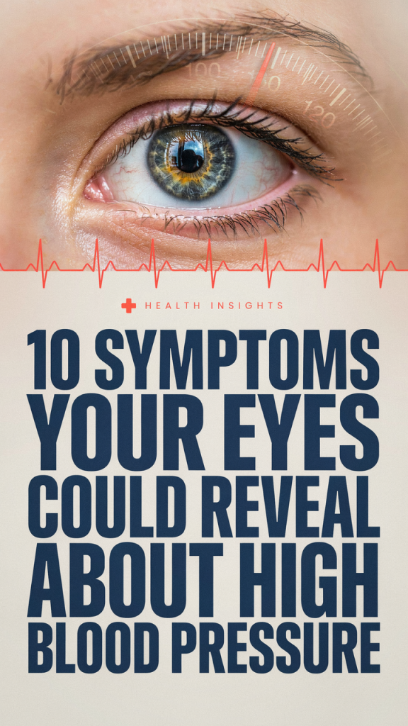
Some of the most telling clues of hypertension are visible only to an eye care professional during a dilated fundus exam or via retinal imaging. These signs help grade the severity of hypertensive retinopathy and guide systemic workup. Below we describe the principal retinal markers and what they mean for overall cardiovascular risk.
Retinal Hemorrhages And Microaneurysms
Retinal hemorrhages appear when fragile capillaries rupture under pressure. They can be flame-shaped (in nerve fiber layer) or dot-blot (in the deeper retinal layers). Microaneurysms are tiny saccular outpouchings of capillaries that indicate weakened vessel walls.
Clinical implications:
- Flame-shaped hemorrhages often reflect superficial nerve fiber layer damage and are common in acute rises of blood pressure.
- Dot-blot hemorrhages and microaneurysms suggest chronic microvascular compromise and are more frequently seen with longstanding hypertension, diabetes, or both.
- The presence of multiple hemorrhages and microaneurysms increases the risk of macular involvement, which directly threatens central vision.
Management focuses on controlling systemic blood pressure and addressing any coexisting vascular disease. Ophthalmic monitoring with fundus photography or OCT helps track progression and detect sight-threatening edema early.
Cotton-Wool Spots And Hard Exudates
Cotton-wool spots are fluffy white patches on the retina caused by localized retinal ischemia, essentially small areas where axoplasmic flow in nerve fiber layer bundles has stalled due to poor perfusion. Hard exudates are yellowish lipid deposits left behind after chronic vascular leakage.
Why they matter:
- Cotton-wool spots are markers of significant microvascular ischemia. Multiple spots suggest widespread retinal hypoperfusion and correlate with higher cardiovascular risk.
- Hard exudates around the macula are concerning because they signal chronic leakage that can impair central vision if it accumulates in the foveal region.
Detecting these lesions should trigger a coordinated plan: tighter blood pressure control, assessment for other vascular risk factors (lipids, glycemic status), and periodic retinal imaging. In advanced cases with macular edema, ophthalmologists may consider intravitreal therapy or focal laser, treatments aimed at preserving central vision while systemic control is optimized.
Papilledema And Optic Nerve Swelling
Papilledema refers to optic disc swelling due to raised intracranial pressure, and while it has multiple causes, malignant or severe uncontrolled hypertension can elevate intracranial pressure enough to produce disc edema. True optic disc swelling may also arise from ischemic optic neuropathy when blood flow to the optic nerve head is compromised.
Signs and symptoms we watch for:
- Blurred disc margins and elevation of the optic nerve head on exam.
- Transient visual obscurations, brief episodes of dimming or graying of vision lasting seconds.
- Associated systemic clues: very high blood pressure readings, headache, nausea, or neurologic deficits.
Papilledema is a medical emergency because it may indicate hypertensive encephalopathy or intracranial hemorrhage. Even without intracranial causes, ischemic optic neuropathy can lead to permanent vision loss. When optic nerve swelling is suspected, we pursue neuroimaging (often MRI) and urgent blood pressure reduction under close supervision.
When To See A Doctor And Emergency Warning Signs
Knowing when to seek care can make the difference between reversible symptoms and permanent vision loss. We err on the side of urgency for several specific presentations.
Seek immediate emergency care (call 911 or go to the ER) if you experience:
- Sudden, painless loss of vision in one or both eyes.
- Sudden onset of a curtain or shadow over part of your vision.
- Severe headache with visual changes, confusion, weakness, or difficulty speaking.
- New double vision with drooping eyelid, slurred speech, or facial weakness.
Make an urgent appointment with your primary care provider or ophthalmologist within 24–48 hours if you notice:
- New persistent floaters or flashes, especially if accompanied by a reduction in visual clarity.
- Recurrent episodes of blurred vision, particularly those that correlate with high blood pressure readings.
- Any new visual field loss or persistent light sensitivity.
What to bring and what clinicians will do:
- Bring a home blood pressure log if you have one: clinic teams often correlate ocular findings with simultaneous blood pressure measurements.
- Eye care professionals will perform a dilated fundus exam, OCT, visual field testing, and fundus photography. In suspected vascular events they may coordinate carotid ultrasound, echocardiography, or neurologic imaging.
- Immediate treatment targets include controlled, monitored lowering of blood pressure in hypertensive emergency, management of ocular complications (e.g., intravitreal therapy for macular edema), and secondary prevention (antiplatelet therapy, statins, anticoagulation when indicated).
Prevention is equally important: regular blood pressure monitoring, adherence to antihypertensive medications, lifestyle measures (sodium reduction, weight management, physical activity), and routine eye exams, especially if we have diabetes or known vascular disease, dramatically reduce the risk of sight-threatening complications.
Conclusion
Our eyes are windows not only to the soul but to our cardiovascular health. In 2026, with better imaging and growing public awareness, we have more opportunity than ever to catch hypertensive damage early. New or sudden visual symptoms, from fluctuating blur and floaters to sudden vision loss or optic nerve swelling, deserve prompt attention because timely intervention can prevent permanent harm. We should keep routine eye exams, consistent blood pressure control, and clear communication with our healthcare team at the top of our prevention strategy. If your eyes start sending unusual signals, don’t wait: act quickly, document what you notice, and get evaluated.