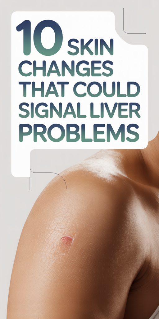
10 Skin Changes That Could Signal Liver Problems
Liver disease often develops quietly, but our skin can act like an early-warning system. In 2026, with better awareness and more accessible diagnostics, recognizing skin signs linked to liver dysfunction can speed diagnosis and improve outcomes. In this text we’ll walk through ten skin changes that commonly, but not exclusively, point to liver problems, explain the underlying mechanisms, and offer practical next steps. We’re not here to replace medical advice, but to help you know what to watch for and when to seek evaluation so liver issues aren’t missed.
How Liver Dysfunction Affects Skin: Key Mechanisms To Know
The liver plays many roles that directly and indirectly affect skin health. When it’s impaired, several biological pathways change and produce visible skin signs. Understanding these mechanisms helps us interpret why particular lesions or symptoms appear.
Bile pigment accumulation
One of the most straightforward links is bile pigment (bilirubin) accumulation. The liver normally conjugates bilirubin and excretes it in bile. When that process is compromised, by hepatocellular injury or cholestasis, bilirubin rises in the bloodstream and deposits in tissues, causing yellowing of the skin and the sclerae (jaundice).
Altered hormone and protein metabolism
The liver metabolizes hormones and synthesizes key proteins, including clotting factors and albumin. Dysfunction can raise estrogen levels (because the liver clears it less efficiently), contributing to vascular changes such as palmar erythema and spider angiomas. Low albumin and clotting factor deficits contribute to easy bruising and petechiae.
Cholestasis and pruritogens
When bile flow is obstructed, pruritogenic substances (bile acids, lysophosphatidic acid, and other mediators) may accumulate and trigger intense itching. This cholestatic pruritus often affects the whole body and can precede jaundice.
Lipid transport and deposition
The liver orchestrates lipid metabolism and clearance of lipoproteins. In conditions where lipid handling is altered, like primary biliary cholangitis or familial hypercholesterolemia, lipid-rich deposits (xanthelasma or xanthomas) can appear on the skin and eyelids, signaling disrupted lipid homeostasis.
Immune dysregulation and skin inflammation
Chronic liver disease can shift immune balance, promoting inflammatory or autoimmune processes that manifest on the skin as rashes, erythema, or autoimmune dermatoses. Some liver diseases are themselves immune-mediated, and skin findings may reflect that systemic activity.
Microvascular and coagulation effects
Liver-related coagulopathy and reduced synthesis of coagulation proteins lead to bleeding tendencies, manifesting as petechiae, easy bruising, or prolonged bleeding from minor trauma. Also, changes in vascular tone from altered hormone metabolism can make superficial vessels more visible.
Putting mechanistic knowledge together helps us prioritize which skin findings are more likely to indicate liver disease and which are nonspecific. Next we’ll look at the specific skin changes clinicians most often associate with hepatic dysfunction and how to interpret them in everyday practice.
Yellowing Of The Skin And Eyes (Jaundice)
Jaundice is the most recognizable cutaneous clue to liver dysfunction, and we should treat it seriously. Clinically, jaundice presents as yellow discoloration of the sclerae (the most sensitive site), skin, and sometimes mucus membranes. The hue correlates with rising bilirubin levels.
What causes it?
Jaundice arises from three broad mechanisms: increased bilirubin production (hemolysis), impaired hepatic uptake/conjugation (hepatocellular disease), or obstruction of bile flow (cholestasis). In liver disease, hepatocellular injury and cholestasis are the usual culprits. Conditions like acute hepatitis, alcoholic liver disease, cirrhosis, and biliary obstruction can all cause jaundice.
How to tell if it’s liver-related
Scleral icterus (yellowing of the whites of the eyes) is an early and reliable sign. If yellowing accompanies dark urine (from conjugated bilirubin being excreted by the kidneys) and pale stools (reduced bile pigment in the gut), cholestasis or obstructive pathology is likely. Systemic symptoms, fatigue, abdominal pain, weight loss, fever, plus abnormal liver blood tests (elevated bilirubin, ALT/AST, alkaline phosphatase, GGT) support a hepatic source.
When to seek care
Because jaundice signals elevated bilirubin and potential hepatic dysfunction, we recommend prompt medical evaluation. Acute onset, associated abdominal pain, fever, mental status changes, or signs of bleeding merit urgent attention. For milder, gradually developing jaundice, outpatient workup with liver enzymes, bilirubin fractionation, and imaging (ultrasound) is usually appropriate.
Common pitfalls
Not all yellowing is jaundice, carotenemia from excess beta-carotene can yellow the skin without scleral involvement. Conversely, mild jaundice may be missed in dark-skinned individuals if we rely only on skin color: scleral examination and lab testing become more important. In short, jaundice remains a high-priority sign that should prompt evaluation for liver disease.
Itching, Rashes, And Persistent Dryness (Pruritus And Cholestatic Skin Changes)
Pruritus, intense itching, is a common and sometimes devastating symptom of liver disease, especially cholestatic disorders. Unlike the itch from dry skin or eczema, cholestatic pruritus can be relentless, worse at night, and resistant to topical moisturizers.
What it looks and feels like
Patients often describe a generalized, deep itch that may come without primary rash. Repeated scratching produces excoriations, weeping, and lichenification (thickened skin). Some people develop secondary bacterial infections from relentless scratching. In cholestasis, the itch may precede other liver symptoms by weeks to months.
Why it happens
Bile acids, endogenous opioids, histamine-independent mediators like lysophosphatidic acid, and increased autotaxin activity have all been implicated as pruritogens in cholestatic liver disease. The exact mechanism is complex and still under study, but it’s clear that systemic accumulation of these substances drives the sensation.
Associated rashes
Certain liver conditions have characteristic cutaneous patterns. For example, primary biliary cholangitis (PBC) often presents with pruritus early in the disease. Viral hepatitis can produce urticarial or maculopapular rashes. Drug-induced liver injury may present with widespread exanthems. We need to consider medication history, recent travel, and systemic signs when evaluating a rash together with pruritus.
Management pointers
Topical emollients and gentle skin care help with dryness but often won’t relieve cholestatic itch. First-line therapies for refractory pruritus include bile acid sequestrants (like cholestyramine), rifampin, or nalfurafine in select regions. In severe cases, bilirubin-lowering interventions or liver transplantation may be required. Because itching can be an early sign, persistent unexplained pruritus should prompt liver function testing.
Vascular Redness And Visible Blood Vessels: Spider Angiomas And Palmar Erythema
Vascular phenomena are classic features of chronic liver disease, particularly cirrhosis. Two findings we commonly see are spider angiomas and palmar erythema. They result from altered hormone metabolism and increased circulating vasodilators.
Spider angiomas
Spider angiomas are central red papules with radiating telangiectatic vessels, often seen on the face, chest, and upper limbs. They occur when elevated circulating estrogens and vasodilatory substances cause dilation of superficial arterioles. While isolated spider angiomas can appear in healthy people (pregnancy, hyperthyroidism), multiple lesions, especially in adults, should raise concern for liver disease.
Palmar erythema
Palmar erythema is characterized by symmetric reddening of the thenar and hypothenar eminences and sometimes fingertips. It’s related to increased vasodilation from higher estrogen and altered nitric oxide metabolism. Like spider angiomas, palmar erythema isn’t specific to the liver (it can occur in rheumatoid arthritis, pregnancy, and thyrotoxicosis), but in context with other signs it supports a hepatic source.
Clinical significance and prognosis
The number and prominence of vascular lesions often correlate with the severity of hepatic dysfunction, though they’re not perfect markers. In cirrhosis, spider angiomas may increase as portal hypertension and estrogen-to-androgen ratios worsen. They’re mostly cosmetic but can help us gauge chronicity and systemic effects of liver disease.
When to act
If we spot multiple spider angiomas or new palmar erythema, especially alongside fatigue, easy bruising, ascites, or abnormal labs, we should pursue liver evaluation. Noninvasive assessments like elastography and standard blood panels can help determine whether portal hypertension or cirrhosis is present.
Fatty Deposits, Bumps, And Yellowish Plaques: Xanthelasma And Xanthomas
Xanthelasma and xanthomas are lipid-rich deposits that appear as yellowish plaques or nodules. They can signal underlying dyslipidemia, cholestatic liver disease, or impaired lipid clearance, conditions that warrant metabolic and hepatic workup.
What we see clinically
Xanthelasma typically shows as soft, yellow plaques on the upper or lower eyelids. Tendinous or eruptive xanthomas appear as firm nodules over tendons or as clusters of papules on the buttocks, elbows, or knees. Eruptive xanthomas are often associated with marked hypertriglyceridemia and can erupt rapidly.
Links to liver disease
Primary biliary cholangitis and other cholestatic disorders can cause secondary hypercholesterolemia because impaired bile excretion disrupts cholesterol clearance. This can lead to xanthelasma and xanthomas. Also, nonalcoholic fatty liver disease (NAFLD) is tightly linked to metabolic syndrome, obesity, insulin resistance, and dyslipidemia, so xanthomas may be a cutaneous clue to broader metabolic-liver pathology.
Differential diagnoses
Not all yellow plaques are xanthelasma. Sebaceous hyperplasia, syringomas, or localized lymphoid aggregates can mimic them. We should consider age, lesion location, lipid profile, and liver tests. Biopsy is rarely needed but can help when the diagnosis is unclear.
Management and implications
Xanthelasma itself is benign and often treated for cosmetic reasons (laser, cryotherapy, excision). But when we encounter these lesions, we should screen for lipid abnormalities and liver dysfunction. Treating underlying dyslipidemia, managing NAFLD, or addressing cholestasis can reduce systemic risk, even if the skin lesions persist.
Bleeding, Petechiae, And Nail Changes (Easy Bruising, Terry Nails, And More)
Coagulopathy and nutritional changes from liver disease produce a spectrum of bleeding and nail findings we can detect on examination. These signs often reflect advanced dysfunction and altered hepatic synthetic capacity.
Easy bruising and petechiae
Reduced synthesis of clotting factors (like fibrinogen, prothrombin, factors VII, IX, X) and thrombocytopenia from hypersplenism or bone marrow suppression can cause easy bruising and petechiae. Petechiae are pinpoint, non-blanching red or purple spots: their presence with mucosal bleeding or prolonged bleeding after minor injuries suggests significant coagulopathy and needs immediate evaluation.
Nail changes: Terry nails and more
Terry nails show whitening of the proximal nail bed with a narrow distal band of normal pink or brown. About one-third of patients with cirrhosis exhibit Terry nails, which correlate with hypoalbuminemia and systemic vascular changes. Other nail findings include Muehrcke’s lines (paired white transverse lines associated with low albumin) and onycholysis in some cases.
Clubbing and other signs
Finger clubbing is not a classic liver sign but can occur in hepatopulmonary syndrome, a complication of advanced liver disease where intrapulmonary vasodilation causes hypoxemia. We should check oxygenation in patients with clubbing and liver disease.
Action steps
When we see bleeding signs or nail abnormalities, we should order a full hepatic panel, coagulation studies (INR, PT, PTT), platelet count, and albumin level. In the setting of coagulopathy, addressing the underlying liver pathology and correcting deficits (vitamin K, plasma transfusion when necessary) becomes urgent, especially before procedures.
Conclusion
Skin findings can be invaluable windows into liver health. From jaundice and cholestatic pruritus to vascular lesions, xanthomas, and bleeding tendencies, these signs may be early clues or markers of advanced disease. We should evaluate persistent, unexplained skin changes, especially when multiple signs cluster together, with liver function testing and appropriate imaging. Early detection improves options and outcomes: treating underlying metabolic issues, stopping hepatotoxic agents, or referring for hepatology assessment can make a real difference. If you spot one of these signs in yourself or a patient, don’t ignore it, use it as a prompt to investigate the liver.